201 Taihe North Road, Baiyun District, Guangzhou, Guangdong Province, China
 020-31922281 \ +8613711194614
-
-
201 Taihe North Road, Baiyun District, Guangzhou, Guangdong Province, China020-31922281 \ +8613711194614


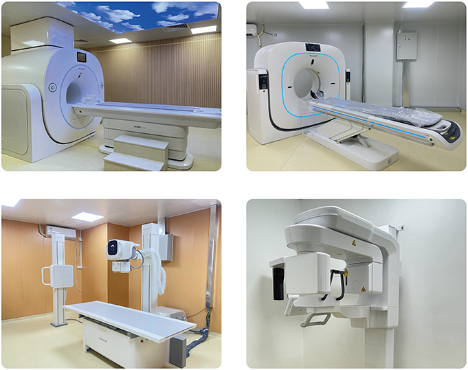
The Imaging Department at Sing May Hospital integrates tumor screening, precise diagnosis, and treatment efficacy evaluation. Equipped with advanced technologies such as DSA, CT, MRI, and DR, the department has established a comprehensive tumor imaging and treatment system centered on “early detection — precise localization — continuous control”:
 Multidimensional Imaging Technology
Utilizes multiphase enhanced CT/MRI scans to accurately detect early and small lesions, clearly defining tumor staging and infiltration extent.
Integrated Interventional Diagnosis and TreatmentPerforms minimally invasive tumor vascular embolization under DSA guidance, achieving seamless integration of “imaging diagnosis and interventional therapy.”
Dynamic Treatment Efficacy MonitoringEmploys radiomics quantitative analysis to monitor real-time responses to radiotherapy, chemotherapy, and targeted therapies, facilitating timely optimization of treatment plans.
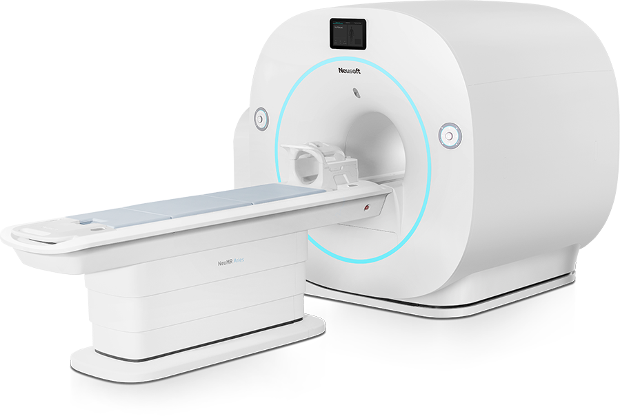
The newly introduced 1.5T magnetic resonance imaging system in our hospital is currently the most advanced 1.5T magnetic resonance imaging equipment in the industry. This equipment adopts a full-process humanized design, making the MRI examination more comfortable and convenient, and comprehensively improving the patients' examination comfort. MRI examination has no X-ray ionizing radiation and is harmless to the human body. It can complete diagnoses that many other imaging examination equipment cannot achieve and has become the most important examination method in imaging examinations.
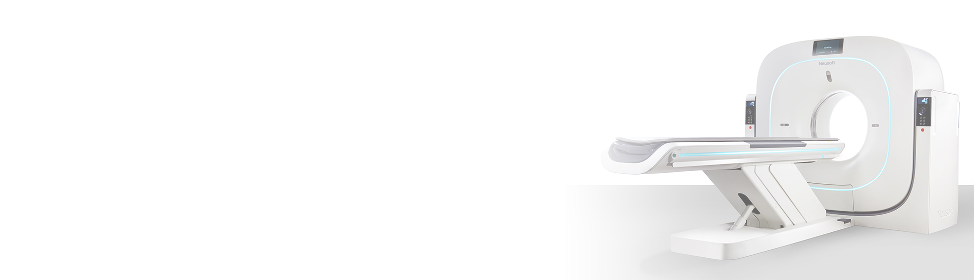
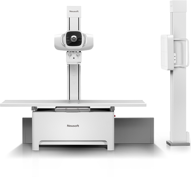
The newly introduced high-end 64-slice 128-row CT in our hospital is an advanced 64-slice 128-row X-ray computed tomography equipment in the current industry. This equipment adopts a full-process humanized design. With extremely short scanning time and very low radiation dose, it can obtain high-definition examination images. At the same time, it has a wide range of applications, almost covering all systems of the whole body. Especially for the detection and diagnosis of lesions in the central nervous system, head and neck, respiratory system, digestive system, urinary system, etc., it has relatively prominent advantages.
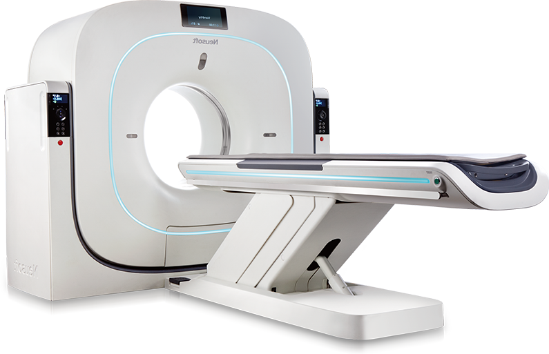
It has advantages such as clear images, fast imaging, ultra-low-dose radiation to patients, and easy access and transmission. For the health screening and disease examination of various parts of the human body, it has very important value in the comprehensive evaluation of tumors in the nervous system, head and neck, abdomen and pelvis, soft tissues, etc., and is an excellent means for disease screening. At the same time, our hospital has introduced an advanced peripheral bone density measurement (DXA) system, which can effectively screen for senile osteoporosis and prevent the occurrence of fragile fractures.
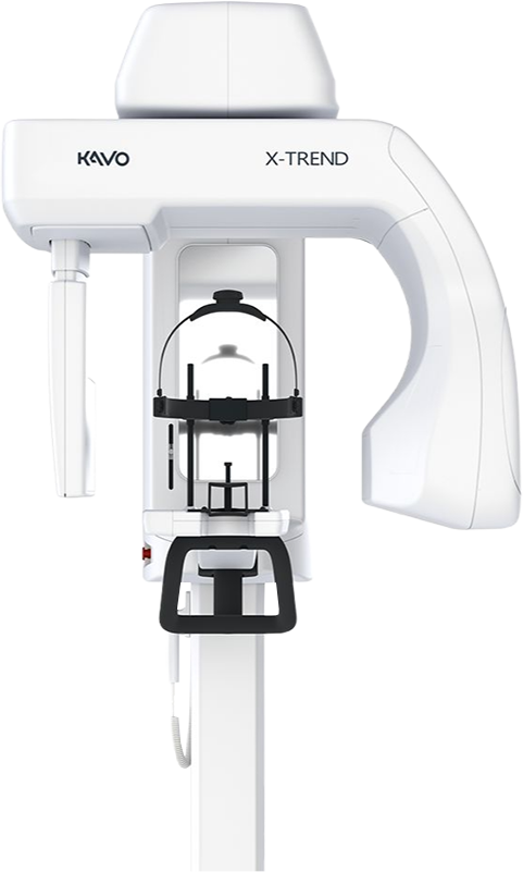
1. Through three-dimensional reconstruction, it can accurately display the lesions of the affected tooth itself and its relationship with surrounding tissues.
2. By performing a 360° rotation scan on the patient, three-dimensional images are obtained, and the affected tooth can be observed from multiple angles and multiple levels, avoiding missed diagnoses caused by image overlap. The diagnosis of root fractures by CBCT is significantly better than that by X-ray periapical films.
3. CBCT can more accurately locate difficult root canals, identify variant root canals, and reduce the possibility of missing root canals, thereby reducing the probability of root canal treatment failure.
4. he sensitivity of CBCT to periapical periodontitis of the affected tooth is higher than that of traditional imaging. Early detection of periapical inflammation and root canal treatment are of great significance for the prognosis.
5. CBCT has relatively prominent advantages in treatments such as orthodontics and dental implants.
 201 Taihe North Road, Baiyun District, Guangzhou, Guangdong Province, China
 020-31922281 \ +8613711194614
 ekoyipsingmay@outlook.com
@Singmay Hospital all rights reserved.