 201 Taihe North Road, Baiyun District, Guangzhou, Guangdong Province, China
 020-31922281 \ +8613711194614
-
-
201 Taihe North Road, Baiyun District, Guangzhou, Guangdong Province, China020-31922281 \ +8613711194614


Personalized and Precision Therapy
In traditional cancer treatment models, standardized methods like surgery, radiotherapy, chemotherapy, and targeted therapy often dominate. However, for patients who are not suitable for surgery or have low physical tolerance, these methods can be limited in effectiveness. To address this, Guangzhou Shengmei Hospital has introduced a personalized, precision-based treatment approach centered on the individual needs of each patient. The hospital is committed to delivering multidisciplinary diagnosis and treatment, comprehensive and standardized personalized cancer care, and specialized rehabilitation programs including hydrogen therapy, natural therapies, and Traditional Chinese Medicine.
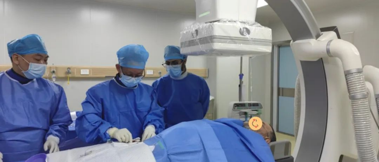
Case Study 1: Interventional Embolization Treatment
1. Patient Background
A patient from Algeria was diagnosed with pancreatic cancer with multiple liver metastases. On January 6, due to upper abdominal pain, he visited a local hospital where chest and abdominal CT scans revealed a mass in the pancreatic tail and multiple nodules in the liver. A liver lesion biopsy indicated a malignancy likely originating from the pancreas. The patient consulted numerous hospitals both domestically and internationally, but was repeatedly denied treatment due to the large tumor size, extensive liver metastases, and complex underlying conditions.

By January 23, the patient experienced worsened upper abdominal and lower back pain, but no fever, nausea, vomiting, chest tightness, or palpitations. Upon recommendation by a friend previously treated at Shengmei, he came to our hospital.
January 23 Enhanced CT findings:
1. Mass in the pancreatic tail, suggestive of pancreatic cancer (Fig. 1, 2)
2. Multiple metastatic liver tumors (Fig. 3)
3. Metastases in the peritoneum and greater omentum
4. Multiple metastases in both lungs
5. Enlarged lymph nodes in the hepatic hilum, retroperitoneum, hepato-gastric space, and right costophrenic angle—likely metastatic


January 24: Under DSA guidance, interventional embolization and intra-arterial chemotherapy were performed for pancreatic cancer with liver metastases. A catheter was inserted into the hepatic artery; angiography showed disorganized tumor vasculature (Fig. 4). A microcatheter was super-selectively advanced into the tumor-feeding artery, and embolization was carried out under DSA (Fig. 5). Post-embolization imaging confirmed effective vascular blockage (Fig. 6).


2. Treatment Plan
The Shengmei medical team selected the most appropriate therapy: DSA-guided tumor embolization.
3. Pre-operative Preparation
Traditional Chinese medicine, physical therapy, and nutritional adjustments were used to stabilize the patient's blood pressure and glucose. Considering religious and cultural differences, a dedicated care team was formed to provide personalized support and dietary accommodations.
4. Embolization Procedure
Angiography revealed clear tumor staining. Physicians super-selectively targeted the tumor’s arterial supply and used embolic microspheres to block the blood flow. Follow-up imaging confirmed that the tumor's blood supply was effectively obstructed.
5. Post-operative Recovery
The patient recovered well and was expected to be discharged and return home within a week.
Case Study 2: Interventional Embolization Treatment
1. Patient Background
“Uncle Li” (alias) visited a hospital in Guangzhou on September 14, 2024, due to pain in the right flank. He was diagnosed with a malignant liver tumor, but received no further anti-tumor treatment. As his upper abdominal pain worsened, he came to Shengmei Hospital for evaluation and treatment.

January 16, 2025 Enhanced CT findings:
1. A mass at the S4/8 junction of the liver, suggestive of hepatocellular carcinoma (HCC)
2. An abnormally enhancing nodule in liver segment S5, suggesting an intrahepatic metastatic lesion


2. Treatment Plan
CT results ruled out surgical contraindications. On January 16, 2025, a CT- and ultrasound-guided liver biopsy was performed, confirming the diagnosis of hepatocellular carcinoma.

January 24, 2025: A DSA-guided embolization was performed for the liver tumor and intrahepatic metastasis with simultaneous injection of chemotherapy drugs. Angiography showed disordered tumor vessels (Fig. 5). A 1.8F microcatheter was super-selectively inserted into the tumor-feeding artery, embolization was completed under DSA, and good lipiodol deposition was observed, indicating successful vascular blockage.


3. Intraoperative Status
The patient's vital signs remained stable during surgery, and he returned to the ward without complications.
About Microvascular Interventional Techniques
In many advanced tumors, nearby large blood vessels or vital organs are invaded, making surgical resection impossible. Vascular interventional therapy offers an alternative: a catheter is inserted into the tumor’s feeding micro-artery, delivering chemotherapeutic agents or embolic materials to starve or kill the tumor directly. This approach is most commonly used for liver tumors.
Under local anesthesia, a fine needle punctures the femoral artery, and a long catheter is threaded through to the tumor site. Medication is administered directly to the tumor area, resulting in a high local drug concentration far exceeding that of systemic chemotherapy, providing a strong anti-tumor effect while minimizing systemic side effects.

HepaSphere Microspheres
These microspheres are normally stored as a powder and expand to 200μm in diameter after absorbing chemotherapy drugs. They are highly stable and can remain inside the tumor for extended periods. In contrast, traditional lipiodol embolic agents are fragile and can break apart under blood flow, potentially obstructing microvessels in normal tissue and causing damage. Moreover, water-soluble chemo drugs quickly separate from lipiodol, limiting sustained drug release and reducing treatment effectiveness.
Once the drug-loaded microspheres block the tumor blood vessels, nutrient supply is cut off, halting further tumor growth. The chemotherapy drugs are then slowly released over two weeks, infiltrating the tumor tissue. Within days, tumor cells begin to apoptose and necrotize.

Case Example: 79-Year-Old Patient with Large Gastric Cancer Liver Metastasis
Treatment Plan: Microsphere embolization using cisplatin 30mg and docetaxel 20mg
 201 Taihe North Road, Baiyun District, Guangzhou, Guangdong Province, China
 020-31922281 \ +8613711194614
 ekoyipsingmay@outlook.com
@Singmay Hospital all rights reserved.